Colon Choir

šŸŽ¶šŸ’© The Choir in Your ColonĀ šŸ’© šŸŽ

This is nothing but mucus to my ears!

Let your colon sing!

And toot a harmony from your butt flute!

iā™”histo

From the musical histologist @Chapman_Histo on Twitter

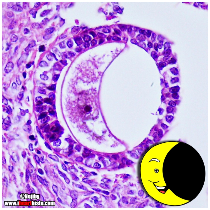
The choir is composed of transverse sections through the crypts of Lieberkuhn that are located in the mucosa of the colon. Thee crypts are intestinal mucus glands. The majority of cells that line them are goblet cells (you see the blue-purple cells interspersed around the singing mouths?) that synthesize and secrete mucus. This mucus is then released into the lumen of the crypt (the open O-mouths) and is squeezed along the gland as the gut contracts and moves. The mucus is eventually spewed out onto the inner lining of the colon. It is here that it acts as a lubricating substance to allow your feces (which have had most of the nutrients and much of the water absorbed form them already – so they are usually fairly dry) smooth passageway into your rectum and along your anal canal when you defecate. This helps prevent damage to the lining of you colon every time you poop and stop things from getting all constipated up in there.

šŸ’©

So next time you have a smooth, contemporary classical movement in C-major – just think about the important role these singing crypts of Leiberkuhn and their goblet cells played in making it happen and rejoice!

šŸ’©


foodorhisto

šŸ½Ā Food or Histology?Ā šŸ½

Some of these slices are edible and some are just slices of dead people.

šŸ…

Which would you put in your mouth and which would you put under your microscope?

šŸ’

Bon appƩtit!

iā™”histo

(Iā€™ll just leave this here. Iā€™ll post the answers soon šŸ™‚ Have fun!)

moon oocyte

šŸŒ™ Crescent Moon Oocytes šŸŒ™

Itā€™s just a phase theyā€™re going through.

iā™”histo

This moon is actually a collapsed oocyte (human egg cell) sitting in a multilaminar primary follicle. These egg-containing follicles are located in the cortex of the female ovary. During the menstrual cycle a handful of follicles that contain these oocytes undergo important structural and functional changes prior to ovulation.

šŸŒ™

The most immature follicle is the primordial follicle. Each month a number of them are kick-started into differentiation. The primordial follicle is identifiable by its circumscribing ring of thin flat epithelial cells. As the follicle matures these cells grow to form a single thick layer of cuboidal cells (unilaminar primary follicle). These follicular cells proliferate further to form a stratified layer of cells (multilaminar primary follicle like those seen here). Some of these primary follicles will develop into secondary follicles. You can identify secondary follicles because their follicular cells have developed fluid filled holes in them (antra) and the follicular cells themselves become known as the stratum granulosum where they play an important role in estrogen production. Eventually the most mature secondary follicles will see all its fluid filled antra merge to form a giant bubble. It is this follicle, the tertiary or Graafian follicle, that houses the oocyte that will be ovulated from the ovary andā€¦ in the presence of some little spermiesā€¦ form a brand new baby human bean.

šŸŒ™

This follicle was shining brightly in the microscope of @Nejiby on Insta

t-rex cells.jpg

šŸ² Dā€™yathinkhesaurus ? šŸ²

The rare histological Dā€™yathinkhesaurus comes out of hiding again in this collection of cells.

Previousy encountered in the now internet famous endometrial tissue section that I posted back here c.2014

iā™”histo

Based on an image seen in the microscope of @pathodaily via Insta

hands

āœ‹ Hands āœ‹

The small bones of the hands developing from a hyaline cartilage scaffold by a process called endochondral ossification.

Histology is from the microscope of @pathologeek via Insta

iā™”histo

Thereā€™s a little devil in all of us

In this case heā€™s in the medullary cavity of the diaphysis of a proximal phalanx.

And heā€™s making work for idle osteons.

iā™”histo

This devil is not an osteon (which is how cortical bone around the outside of a bone is organized) but a trabecula, which is how the spongy bone within the diaphysis of a long bone is organized.Ā 

His peepers are mature bone cells (osteocytes) residing in white lacunae.Ā 

His smile is a bony lamellar.Ā 

His horns are remnants of the endosteum and some of the hematopoietic cells (blood forming cells) that occupy the marrow cavity (white space in this image).

Stewie Griffin or a Fecal Parasite

ā€œHello, mother.Ā 

I come bearing a gift.Ā 

I’ll give you a hint.Ā 

It’s in my diaper and it’s not a toaster.ā€

iā™”histo

Stewieā€™s head is the two-cell stage (infective) egg of a small parasitic roundworm called whipworm (Trichuris trichuria).Ā 

It is ready to make its way into your intestines and after about 3 months will hatch into a whipworm. Once mature, the whipworm burrows her head and mouth parts into the delicate and comfortable mucosa of your large intestine to feed. It is here that she will trigger an inflammatory response and causes bouts of abdominal pain with plenty of (sometimes bloody) diarrhea. A disease called trichuriasis.Ā 

Meanwhile, her vulva- containing end, dangles into the lumen of the intestine and has sex with as many nearby male worms as possible.

Our whipworm now becomes a momma by firing out somewhere between 2,000-10,000 eggs….

Everyday!Ā 

This life of feeding and egg-laying, if left untreated, can last for the lifespan of a whipworm which can be around 5 years… thatā€™s a very busy worm and lots of eggs.

Her Stewie-headed eggs get flushed out of your body (with all your bloody diarrhea). Depending on where you live (roundworms are most common in tropical, developing countries with poor sanitation), these eggs can make their way into the water supply or food supply where they can be ingested once again and the whole process starts over.

Now watch me whipworm. Now watch me nae nae.