🍴 Fed or Dead? 💀 [ANSWERS]

Some of these slices are edible and some of these slices are just dead people.

Which would you put in your mouth and which would you put under your microscope? 

As promised here are the answers to this week’s pathological dilemma. 

Congratulations to everyone who avoided an embarrassing, cannibalistic faux pas at the dining table. To the rest of you – don’t be surprised if your friends suddenly find themselves “busy” when you next host a dinner party.

Fed or Dead

1. 🍴 Fed 🍴 

This is red cabbage delicious pickled or in slaw. 

2. 💀 Dead 💀

This is a slice through your sciatic nerve. Great for contracting your hamstrings (also not food) but not so tasty on a sandwich. The nerve is composed of the axons of many neurons all cut in half so they look like hundreds of little o’s filling the specimen. The whole nerve is surrounded by a connective tissue perineurium.

3. 💀 Dead 💀 

This is a slice through the jejunum of the small intestine. Great for digestion or indigestion depending on whether you choose munch or microscope. (Although technically the outer wall of the intestines are what they use for the skin of sausages – so technically this could go both ways). You can tell it’s small intestine because all those projections are villi. They increase the surface area of the lining of your small intestine to improve its efficiency at absorbing all the nutrients from the food you eat. You can tell it is jejunum over the other regions of the small intestine because it doesn’t have Brunner’s glands (duodenum) or Peyer’s patches (ileum).
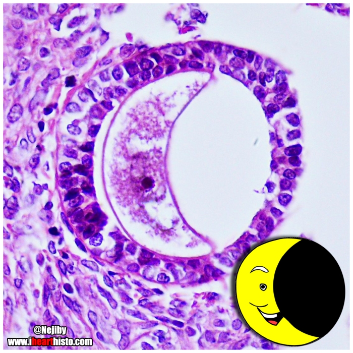
4. 💀 Dead 💀 

This is an ovary. Think of a baby not a Baby Ruth. All those little ‘holes’ in this tissue are ovarian follicles at different stages of development. Each contains a tiny egg (also not food). One of these eggs will eventually be ovulated and under the right conditions* has the potential to form a real life baby (*spermatozoa required).

5. 🍴 Fed 🍴 

Sweet purple potato. All the yummyness of a traditional sweet potato but with an H&E hue.

6. 💀 Dead 💀 

This is a Pacinian corpuscle in the dermis of the skin. Not for pressure cooking but great for sensing pressure. They are nerve endings that are sensitive to vibration and sudden disturbances in pressure, such as when feeling a rough v smooth surface or grasping/releasing an object.

7. 🍴 Fed 🍴 

This is the beet root. A staple in borscht. Warning: Eat too much and it will turn your poop red.

8. 🍴 Fed 🍴 

Dragon fruit. The red variety of the pitaya. They look a bit like a disembodied testicle in cross-section and have a disappointing taste for such a vibrant looking fruit. 

9. 🍴 Fed 🍴 

Red onion. Perfect in that summer salad or in an onion jam!

Bon appétit!

i♡histo

Colon Choir

🎶💩 The Choir in Your Colon 💩 🎶

This is nothing but mucus to my ears!

Let your colon sing!

And toot a harmony from your butt flute!

i♡histo

From the musical histologist @Chapman_Histo on Twitter

The choir is composed of transverse sections through the crypts of Lieberkuhn that are located in the mucosa of the colon. Thee crypts are intestinal mucus glands. The majority of cells that line them are goblet cells (you see the blue-purple cells interspersed around the singing mouths?) that synthesize and secrete mucus. This mucus is then released into the lumen of the crypt (the open O-mouths) and is squeezed along the gland as the gut contracts and moves. The mucus is eventually spewed out onto the inner lining of the colon. It is here that it acts as a lubricating substance to allow your feces (which have had most of the nutrients and much of the water absorbed form them already – so they are usually fairly dry) smooth passageway into your rectum and along your anal canal when you defecate. This helps prevent damage to the lining of you colon every time you poop and stop things from getting all constipated up in there.

💩

So next time you have a smooth, contemporary classical movement in C-major – just think about the important role these singing crypts of Leiberkuhn and their goblet cells played in making it happen and rejoice!

💩


foodorhisto

🍽 Food or Histology? 🍽

Some of these slices are edible and some are just slices of dead people.

🍅

Which would you put in your mouth and which would you put under your microscope?

🍒

Bon appétit!

i♡histo

(I’ll just leave this here. I’ll post the answers soon 🙂 Have fun!)

teratomaaackbar

🌠 Admiral Ackbar Teratoma 🌠

“It’s a trap!”

An intramuscular tumor leading a rebellion against an embryological Empire.

i♡histo
I Heart Histo – Histology
www.ihearthisto.com

🚀

A teratoma is a tumor that contains numerous different tissues from different locations in the body. For example a brain tumor that is discovered to contain teeth, hair and bone may be a teratoma. It may seem strange but it is amazing that it doesn’t happen more often if you think about where all of our tissues actually come from… During embryological development our entire body develops from cells that arise from only three different germ cell layers (endoderm, ectoderm and mesoderm). These cells will differentiate into whatever tissue they are told to by different signalling molecules. Remarkably this happens accurately 99.9% of the time but in some cases, the cells might get confused and turn into the wrong tissue in the wrong location resulting in a teratoma. The teratoma shown here is within a skeletal muscle near your shin bone (the tibialis anterior muscle for the anatomists out there). It contains, cartilage, bone, blood vessels, skin and glands.

🚀

Seen in the microscope of @jkbiologic on Insta

moon oocyte

🌙 Crescent Moon Oocytes 🌙

It’s just a phase they’re going through.

i♡histo

This moon is actually a collapsed oocyte (human egg cell) sitting in a multilaminar primary follicle. These egg-containing follicles are located in the cortex of the female ovary. During the menstrual cycle a handful of follicles that contain these oocytes undergo important structural and functional changes prior to ovulation.

🌙

The most immature follicle is the primordial follicle. Each month a number of them are kick-started into differentiation. The primordial follicle is identifiable by its circumscribing ring of thin flat epithelial cells. As the follicle matures these cells grow to form a single thick layer of cuboidal cells (unilaminar primary follicle). These follicular cells proliferate further to form a stratified layer of cells (multilaminar primary follicle like those seen here). Some of these primary follicles will develop into secondary follicles. You can identify secondary follicles because their follicular cells have developed fluid filled holes in them (antra) and the follicular cells themselves become known as the stratum granulosum where they play an important role in estrogen production. Eventually the most mature secondary follicles will see all its fluid filled antra merge to form a giant bubble. It is this follicle, the tertiary or Graafian follicle, that houses the oocyte that will be ovulated from the ovary and… in the presence of some little spermies… form a brand new baby human bean.

🌙

This follicle was shining brightly in the microscope of @Nejiby on Insta

t-rex cells.jpg

🐲 D’yathinkhesaurus ? 🐲

The rare histological D’yathinkhesaurus comes out of hiding again in this collection of cells.

Previousy encountered in the now internet famous endometrial tissue section that I posted back here c.2014

i♡histo

Based on an image seen in the microscope of @pathodaily via Insta


Pokémon Histology: Gotta catch ‘em all!


1. Mantyke in a cervico-vaginal smear

Obtained during a Pap smear of a patient treated for cervical cancer. The epithelial cells are abnormal showing irradiation effects

2. A Gastly diagnosis
A case of acute megakaryoblastic leukemia – a type of leukemia involving the bone marrow cells that are precursors to the cells that form platelets (megakaryocytes).

3. Mespirit during embryonic neurulation
This image shows a transverse section through an embryo during the process of neurulation – formation of the central nervous system. It has not yet completed because the neural tube (top of Mespirit’s head) is not yet closed it is still open on the dorsal (back) surface of the embryo (U-shaped) rather than a sealed tube (O-shape).

i♡histo

http://www.ihearthisto.com